Researchers: Zengbo Wang (Bangor University), John Thomas (Bangor University), Yukun Lai (Cardiff University), Alan Parker (Cardiff University)
The physical law of light diffraction means that optical microscopes cannot detect objects with diameters smaller than ~200 nm, including most viruses. By drawing together their expertise from a range of disciplines, this team of researchers from Welsh Crucible 2015 developed a new type of microsphere and microcylinder-based optical superlens that can perform large-area scanning super-resolution imaging of moving samples at resolution of about 100 nm. Live virus and nano-samples that were previously ‘invisible’ under microscope are now clearly revealed under the new superlens.
The project involved interdisciplinary joint efforts from an optical engineer (Zengbo Wang) who designed the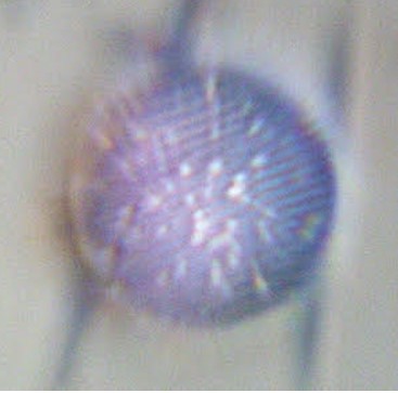 superlens and made the prototype, a scanning expert (John Thomas) who developed the scanning strategy, a computer scientist (Yukun Lai) who made be-spoke software for imaging capture and stitching, and finally a virologist (Alan Parker) who grew live virus for the imaging experiments in the project.
This work laid down the foundation for their more recent work on the development of more advanced superlens technologies, including the “spider silk superlens” and the “nanoparticle superlens”, which attracted lots of media attention. Zengbo Wang is leading the development of a new microfluidic superlens device for biological live cells and viruses imaging for an international EU Horizon project researching semiconductor-based ultrawideband micromanipulation of cancer stem cells.